 繁體中文
繁體中文  English
 簡體中文
簡體中文 日本語
日本語
代理品牌
superGolgi Kit
superGolgi Kit 為 Bioenno lifescience 的主打產品
每年都有相當多的文獻指名用Bioenno superGolgi kit
染色的效果絕佳,即使是不好染色的海馬迴,也可以染出清晰的成果
產品規格:12 Blocks (~1×1×2 cm) of brain tissue
保存期限:18 Months

The superGolgi Kit, from Bioenno, provides an enhanced Golgi-Cox staining system. superGolgi is designed for the staining and impregnation of dendrites and dendritic spines of neurons. It is based on the principle of Golgi-Cox impregnation. The Kit has been extensively tested on various brain tissues including those harvested from rats, mice, cats, rabbits, monkeys, as well as postmortem brains of humans. See The Golgi Method and superGolgi Kit – An Overview and Introduction to the Golgi Method for a technical overview about this technique and its uses in the neurohistology field.
The superGolgi Kit yields both stable and high quality labeling of dendrites and spines. The impregnation time takes 7 to 14 days depending on the age and size of tissues. The Kit can be stored in a dark area at room temperature (22 +/- 2 deg C) for up to 18 months.
Product Features
- Reliable and high contrast impregnation and staining of dendrites and dendritic spines
- Suitable for freshly harvested brain tissues
- Extensively tested on a broad range of species
- Employs a refined and stable Golgi-Cox solution
- Impregnation time of 1 to 2 weeks
- Streamlined staining protocol
- Sufficient for 10-12 blocks (~1 x 1 x 2 cm) of brain tissue
- For in-vitro lab use
- Warranty: 18 months
Proven Results
Dendritic branches and spines have been reliably stained and impregnated with the superGolgi Kit.
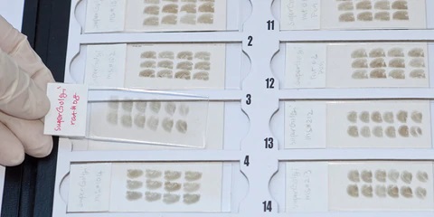
Fig. 1 – Impregnated and Stained Brain Sections
Mounted on Adhesive Glass Slides
As shown in Fig. 1, following the superGolgi Kit protocol, brain tissues from a rat and mouse were impregnated, sliced, and stained. The stained sections can be stored at room temperature.
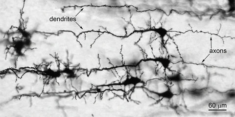
Fig. 2 – Impregnated and Stained Pyramidal Neurons in the Cortex
As shown in Fig. 2, the superGolgi Kit was used to impregnate and stain the pyramidal neurons in the neocortex of a postnatal day 2 (P2), C57BL mouse (20x objective lens). Dendrites and axons were well impregnated and stained.
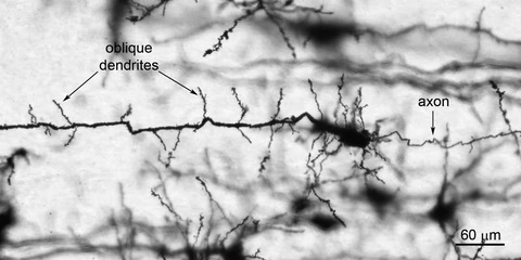
Fig. 3 – Pyramidal Neuron in the Hippocampal CA1 Area
As shown in Fig. 3, the superGolgi Kit was used to impregnate and stain the pyramidal neuron in the CA1 area of the hippocampus of a P2 C57BL mouse (20x objective).
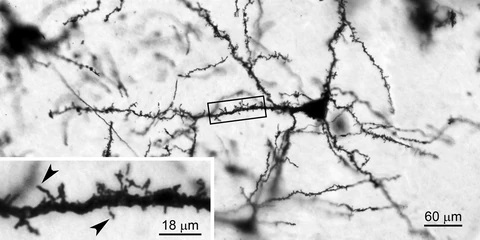
Fig. 4 – Pyramidal Neuron in the Hippocampal CA3 Area
As shown in Fig. 4, the superGolgi Kit was used to impregnate and stain the pyramidal neuron in the CA3 area of the hippocampus of a P7 C57BL mouse (20x objective). The boxed area was magnified to show the filopodia-like protrusions (arrowheads), which are the immature dendritic spines.
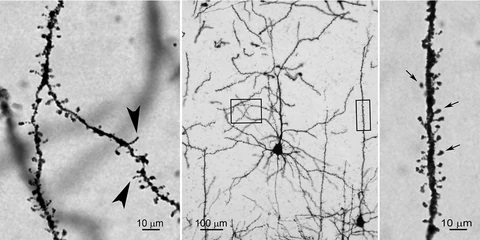
Fig. 5 – Pyramidal Neurons and Labeled Dendritic Protrusions
As shown in Fig. 5, pyramidal neurons were stained using the superGolgi Kit. The pyramidal neurons were taken from the frontoparietal cortex (motor area) of a P21 CD1 mouse. The boxed areas in the Middle panel, shown at 20x, were magnified to 100x and presented in the Left and Right panels. On the Left panel, oblique branches are shown. Arrowhead-indicated, filopodia-like protrusions, which are the immature dendritic spines, are often observed at this age. On the Right panel, mature dendritic spines on the main branch are indicated by arrows.
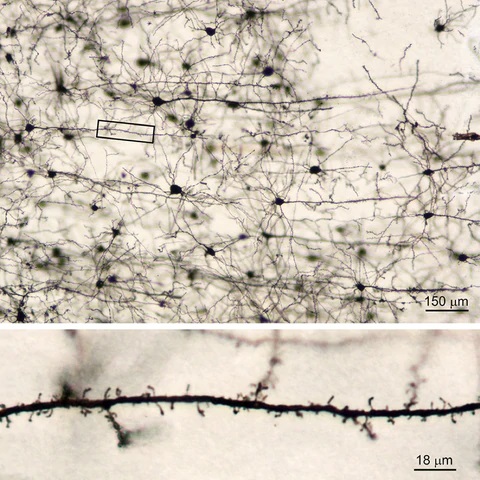
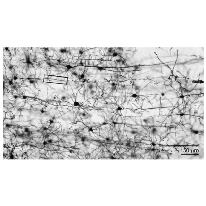
Fig. 6 – Impregnated Cortical Neurons and Labeled Dendritic Protrusions
As shown in Fig. 6, cortical neurons were stained using the superGolgi Kit. These neurons were taken from the frontoparietal cortex (somatosensory area) of a P30 C57BL mouse. The boxed area in the Top panel, shown at 10x, was magnified (63x objective) to highlight the dendritic protrusions.
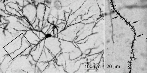
Fig. 7, Left and Right – A Striatal Neuron and Labeled Dendritic Spines (denoted by arrows)
As shown in Fig. 7, the superGolgi Kit was used to impregnate and stain the striatal neuron which was taken from the posterior caudate of a 2-month old Wistar rat. (Left Panel: 20x objective; Right Panel: Boxed area was magnified to 63x)
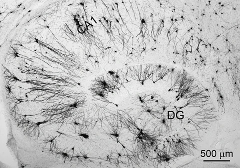
Fig. 8 – Impregnated and Stained Hippocampus
As shown in Fig. 8, the superGolgi Kit was used to impregnate and stain the hippocampal neurons of a 5-month old C57BL mouse (4x objective lens). “DG” denotes dentate gyrus.
Importance of Golgi-Cox Impregnation and Staining
Golgi-Cox impregnation and staining allows scientists to clearly visualize the soma, dendrites, and dendritic spines of neurons from the brain tissues of various animals including rats, mice, cats, rabbits, and monkeys, as well as tissues obtained from post-mortem human brains. Understanding the morphology of these cellular structures is critical for research on the effects of various diseases on the brain including Alzheimer’s. In addition, understanding these structures allows scientists to better understand the effects of aging and the effects of man-made toxins and illicit drugs (e.g. methamphetamine) on the brain. Furthermore, Golgi-Cox impregnation and staining allows scientists to further understand neuroplasticity and neuroprotection.
Importance of Dendrites of Neurons
Dendrites of neurons make up 95% of the total volume of the neuron. Synapses, which are structures that permit neurons to pass electrical and chemical signals to another cell, are found on dendrites. If there are any changes to the dendrites, this will cause neuron damage. It is important to characterize these dendrites to determine if there is any branching atrophy or spine loss.
2017
-
5-Fluorouracil chemotherapy upregulates cytokines and alters hippocampal dendritic complexity in aged mice
Thomas R. Grovesa, Ryan Farrisd, Julie E. Andersona, Tyler C. Alexandera, Frederico Kiffera, Gwendolyn Cartera, Jing Wanga, Marjan Boermaa, Antiño R. Allen(2017), Behavioural Brain Research,316, 215–224
Link: http://www.sciencedirect.com/science/article/pii/S0166432816305575
2016
-
Neuropsin Inactivation Has Protective Effects against Depressive-Like Behaviours and Memory Impairment Induced by Chronic Stress
Simon Chang, Philane Bok, Cheng-Pu Sun, Andrew Edwards, Guo-Jen Huang(2016), PLoS Genet, 12 (10), e1006356
Link:http://journals.plos.org/plosgenetics/article?id=10.1371/journal.pgen.1006356 -
Contrasting the effects of proton irradiation on dendritic complexity of subiculum neurons in wild type and MCAT mice
Nicole N. Chmielewski, Chongshan Caressi, Erich Giedzinski, Vipan K. Parihar, Charles L. Limoli(2016), Environmental and Molecular Mutagenesis, 57, 364-371
Link:http://onlinelibrary.wiley.com/doi/10.1002/em.22006/full -
Postnatal Gene Therapy Improves Spatial Learning Despite the Presence of Neuronal Ectopia in a Model of Neuronal Migration Disorder
Huaiyu Hu, Yu Liu, Kevin Bampoe, Yonglin He and Miao Yu(2016), Genes, 7(12), 105
Link:http://www.mdpi.com/2073-4425/7/12/105 -
Adult microbiota-deficient mice have distinct dendritic morphological changes: differential effects in the amygdala and hippocampus
Pauline Luczynski, Sean O. Whelan, Colette O’Sullivan, Gerard Clarke, Fergus Shanahan, Timothy G. Dinan and John F. Cryan(2016), European Journal of Neuroscience, 44, 2654–2666
Link: http://onlinelibrary.wiley.com/doi/10.1111/ejn.13291/pdf -
Cranial grafting of stem cell-derived microvesicles improves cognition and reduces neuropathology in the irradiated brain
Janet E. Baulcha, Munjal M. Acharyaa, Barrett D. Allena , Ning Rua , Nicole N. Chmielewskia , Vahan Martirosiana , Erich Giedzinskia , Amber Syagea , Audrey L. Parka , Sarah N. Benkea , Vipan K. Parihara , and Charles L. Limolia (2016). PNAS, 113 (17), 4836–4841.
Link: http://www.pnas.org/content/113/17/4836.full -
RBM4 promotes neuronal differentiation and neurite outgrowth by modulating Numb isoform expression
Woan-Yuh Tarna, Hung-Che Kuoa, Hsin-I. Yua, Shin-Wu Liua, Ching-Tzu Tsenga, Dodda Dhananjayaa, Kuan-Yang Hunga, Chi-Chiang Tua, Shuo-Hsiu Changa, Guo-Jen Huangb, and Ing-Ming Chiuc (2016). Molecular Biology of the Cell, 27(10),1676-83.
Link: http://www.ncbi.nlm.nih.gov/pubmed/27009199 -
Combined Treatment With Environmental Enrichment and (-)-Epigallocatechin-3-Gallate Ameliorates Learning Deficits and Hippocampal Alterations in a Mouse Model of Down Syndrome
Silvina Catuara-Solarz, Jose Espinosa-Carrasco, Ionas Erb, Klaus Langohr, Juan Ramon Gonzalez, Cedric Notredame and Mara Dierssen(2016). eNeuro, 3(5), 0103-16.
Link: http://eneuro.org/content/3/5/ENEURO.0103-16.2016
2015
-
Targeted overexpression of mitochondrial catalase prevents radiation-induced cognitive dysfunction
Parihar, V. K., Allen, B. D., Tran, K. K., Chmielewski, N. N., Craver, B. M., Martirosian, V., … & Limoli, C. L. (2015). Antioxidants & Redox Signaling, 22(1), 78-91.
Link: http://www.ncbi.nlm.nih.gov/pubmed/24949841 -
Stem Cell Transplantation Reverses Chemotherapy-Induced Cognitive Dysfunction
Munjal M. Acharya, Vahan Martirosian, Nicole N. Chmielewski, Nevine Hanna, Katherine K. Tran, Alicia C. Liao, Lori-Ann Christie, Vipan K. Parihar, and Charles L. Limoli (2015), Cancer Research, 75, 676-686.
Link: http://www.ncbi.nlm.nih.gov/pubmed/25687405 -
Short-term modern life-like stress exacerbates AB-pathology and synapse loss in 3xTg-AD mice
David Bagliett-Vargas, Yuncai Chen, Dongjin Suh, Rahasoon R. Ager, Carlos J. Rodriguez-Ortiz, Rodrigo Medeiros, Kristoffer Myczek, Kim N. green, Tallie Z. Baram, Frank M. LaFerla (2015). Journal of Neurochemistry, 134, 915-926.
Link:http://www.ncbi.nlm.nih.gov/pubmed/26077803 -
Human endogenous retrovirus-K contributes to motor neuron disease.
Wenxue Li, Myoung-Hwa Lee, Lisa Henderson, Richa Tyagi, Muzna Bachani, Joseph Steiner, Emilie Campanac, Dax A. Hoffman, Gloria von Geldern, Kory Johnson, Dragan Maric, H. Douglas Morris, Margaret Lentz, Katherine Pak, Andrew Mammen, Lyle Ostrow, Jeffrey Rothstein and Avindra Nath (2015). Science Transitional Medicine, 7, 307, 307ra153.
Link:http://stm.sciencemag.org/content/7/307/307ra153.full-text.pdf+html
2014
-
Synergistic effects of amyloid-beta and wild-type human tau on dendritic spine loss in a floxed double transgenic model of Alzheimer’s disease.
Chabrier, M. A., Cheng, D., Castello, N. A., Green, K. N., & LaFerla, F. M. (2014). Neurobiology of Disease, 64, 107-117.
Link: http://www.ncbi.nlm.nih.gov/pubmed/24440055